

San Diego Dental Health Center UCSD
Periapical Radiolucencies with Radiopacities
Training details
US$299
Details and registration continue in the Dentoflow application.
About this training
Content
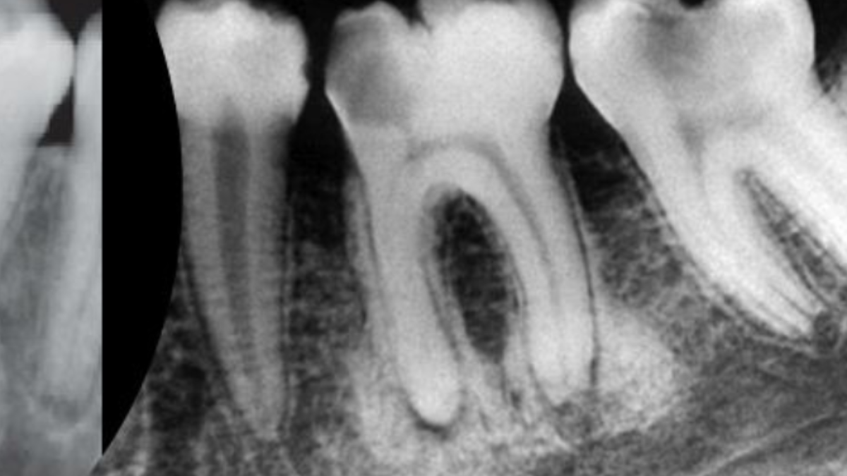
Great attention has been given to the study of radiolucent periapical lesions to avert possible misdiagnosis of apical periodontitis associated with certain non-endodontic lesions. There are a significant number of radiopaque lesions found in the periapical region, which could be equally relevant to dental practices. The diagnosis and management of these radiopaque lesions could be challenging to the dentist or dental specialist. These bone alterations could be neoplastic, dysplastic or of metabolic origin. In the context of the more widespread use of cone-beam CT, a detailed review of the radiological appearance of radiopaque inflammatory and non-inflammatory lesions is timely and may aid dentist perform a differential diagnosis of these lesions. Distinguishing between inflammatory and non-inflammatory lesions simplifies diagnosis and consequently aids in choosing the correct therapeutic regimen. This course discusses the radiographic appearance on 2D radiograph and on CBCT, the pathophysiology and management aspects of radiopaque lesions, and illustrates the differential diagnoses of these lesions.
What you'll learn
Learn the most common differential diagnosis of radiopaque lesions found in the periapical region
Learn the most common differential diagnosis and pathophysiology of the mixed density lesions
Get familiar with the treatment and management of these lesions
Goals
There are a significant number of radiopaque lesions found in the periapical region, which could be equally relevant to dental practices. The diagnosis and management of these radiopaque lesions could be challenging to the dentist or dental specialist. These bone alterations could be neoplastic, dysplastic or of metabolic origin. In the context of the more widespread use of cone-beam CT, a detailed review of the radiological appearance of radiopaque inflammatory and non-inflammatory lesions is timely and may aid dentist perform a differential diagnosis of these lesions.
Prerequisites
no requirements